2023 FDA Science Forum
Assessment of trabecular bone stiffness using radiomics and deep-learning features
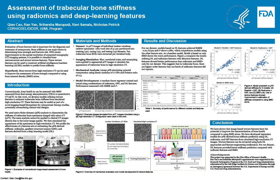
- Authors:
- Center:
-
Contributing OfficeCenter for Devices and Radiological Health
Abstract
Introduction: Evaluation of bone fracture risk is important for the diagnosis and treatment of osteoporosis. Bone stiffness is one major factor in determining bone strength and fracture risk. With recent improvements in the spatial resolution of computed tomography (CT) imaging systems, it is possible to visualize bone microstructure and extract texture features. These texture features can be used to construct artificial intelligence/machine learning (AI/ML) models to predict bone stiffness. We hypothesize that bone texture can be used to improve the assessment of bone strength compared to using bone mineral density (BMD) alone. Purpose: In this work, we develop models utilizing deep learning (DL) features, radiomics features, and gradient structure tensors (GSTs) to estimate trabecular bone stiffness from high-resolution CT. These features may be useful as part of a novel imaging-based biomarkers for osteoporosis therapy studies, potentially streamlining clinical trial designs. Methods: Our dataset contains micro-CT images of 70 individual lumbar vertebrae from patients. Ten trabecular bone ROIs were extracted from each vertebral body and their structural anatomy was segmented. The mechanical stiffness of each ROI was estimated using micro-finite element (µFE) analysis. Blur and correlated noise derived from clinical high-resolution CT systems were then added to the trabecular bone ROIs to generate simulated CT images. A 3D residual DL network (ResNet) was trained to extract DL features to predict µFE-derived bone stiffness from the simulated CT images. Radiomics and GST features of bone ROIs were also computed for the same task. Results: The prediction results for DL features, radiomics, and GST features combined showed the best performance with a root mean square error (RMSE) of 2.646 N/μm and an R2 of 0.881. The performance of DL features alone was superior to BMD alone or using radiomic features alone. Additionally, incorporating orientation information into the models via GST features resulted in improved performance. Conclusion: We demonstrated that µFE-estimated mechanical properties of lumbar vertebral trabecular bone can be inferred from high-resolution CT images and that a combination of DL, radiomic, and GST features provides the highest risk prediction performance, suggesting that an improved texture-based imaging biomarker for facture risk in osteoporosis studies may be possible.