2021 FDA Science Forum
Radiomics Model for Bone Fragility Assessment from Radiographic Projection Images
- Authors:
- Center:
-
Contributing OfficeCenter for Devices and Radiological Health
Abstract
Introduction
Assessment of bone fragility is key for staging and management of osteoporosis. Currently, bone mineral density (BMD) is used for evaluating fracture risk and can be measured by quantitative CT or dual-energy x-ray absorptiometry (DXA). Fragility depends on both the mineral distribution and bone architecture. Therefore, recent efforts have emphasized characterization of bone microarchitecture. In this Office of Women’s Health (OWH) project, we focus on characterizing bone fragility from x-ray projection images. Projection-based modalities may be beneficial because they are commonly available and may be used to determine bone fragility without a high-resolution 3D representation of the full bone microstructure.
Purpose
The overall goal of our research effort is to investigate novel imaging-based biomarkers for osteoporosis therapy studies such that a more streamlined trial design may be possible. The specific objective of this study is to develop an artificial intelligence/machine learning (AI/ML) radiomics model to predict the risk of bone failure from radiographic projection images.
Methods
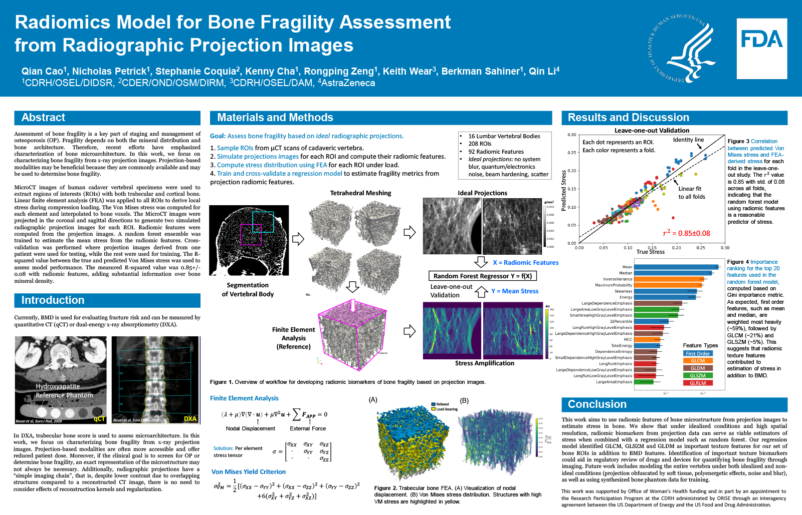
MicroCT images of 16 human cadaver vertebral specimens were used to extract 208 regions of interest (ROIs). Linear finite element analysis was applied to all ROIs to derive local stress during compression loading. The equivalent Von Mises stress was computed for each element and interpolated to bone voxels. Von Mises stress amplification, defined as the Von Mises stress of the voxel divided by uniaxial stress experienced by the ROI, was calculated. The MicroCT images were projected in the coronal and sagittal directions to generate two simulated radiographic projection images for each ROI. Radiomic features (92 features total) were computed from the projection images. A random forest ensemble was trained to estimate the mean stress amplification from the radiomic features. Cross-validation was performed where projection images derived from one patient were used for testing, while the rest were used for training. The R-squared value between the true and predicted Von Mises stress amplification was used to assess model performance.
Results
The measured R-squared value was 0.85+-0.08 indicating the radiomic features added substantial information over bone volume fraction.
Conclusion
Radiomic features from projection images can be combined with nonlinear regression techniques to predict bone fragility.