2021 FDA Science Forum
Effects of Silver Nanoparticles on Endothelial Cells under Physiologically Relevant Hemodynamic Conditions: Increased Cytotoxicity and Inflammation
- Authors:
- Center:
-
Contributing OfficeCenter for Devices and Radiological Health
Abstract
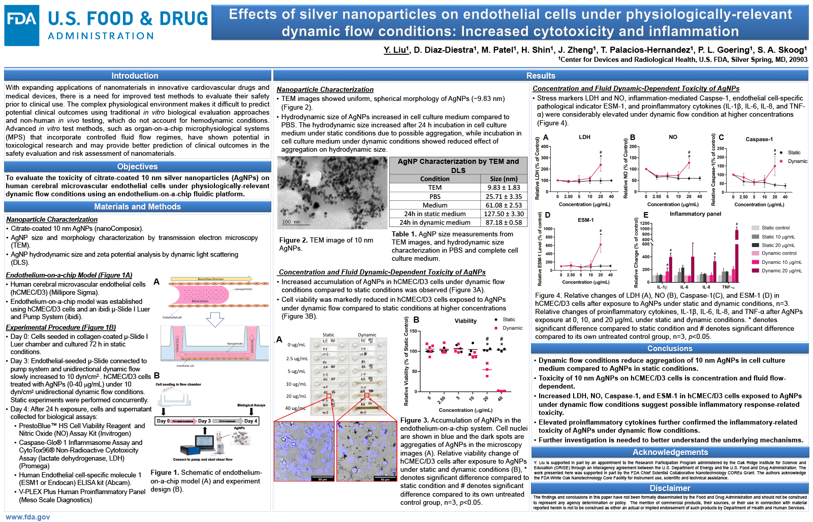
The toxicity of nanomaterials depends on their physicochemical properties as well as the physiological environment. However, the complexities of the physiological environment make it very difficult to predict the potential clinical outcomes using data from traditional in vitro biological evaluation approaches and non-human in vivo testing. As part of the global efforts for reducing animal testing, advanced in vitro test methods such as organ-on-a-chip microphysiological systems (MPS) have shown potential in toxicological research by providing a more physiologically-relevant microenvironment. In this study, a microfluidic system was adapted to establish an endothelium-on-a-chip model for investigating the effects of silver nanoparticles (AgNPs) on endothelial cells under physiologically-relevant hemodynamic condition. Human cerebral microvascular endothelial cells (hCMEC) and 10 nm AgNPs were used in the endothelium-on-a-chip model based on initial preliminary screening of different sized AgNPs across four types of human endothelial cell lines. hCMEC were cultured to confluency on the platform under static (no-flow) conditions and then exposed to AgNPs at concentrations ranging from 0 to 40 ?g/mL under 10 dyn/cm2 flow conditions. In parallel, the same treatments of AgNPs were performed on cells in static culture. After 24-hour AgNP exposure, hCMEC exhibited significantly higher toxicity under flow conditions compared to cells under static conditions. hCMEC viability decreased over 50% and elevated lactate dehydrogenase levels under flow conditions after treatment with 20 ?g/mL and 10-20 ?g/mL AgNP, respectively. Nitric oxide levels were also substantially increased after AgNP treatment under flow conditions. Caspase-1, an inflammatory response initiator, and endocan (ESM-1), an endothelial-specific inflammatory response marker, increased by 50% and over 6-fold increase respectively, after treatment with 20 ?g/mL AgNPs, suggesting possible inflammatory response-related toxicity. These findings demonstrate the importance of physiologically-relevant environments in nanomaterial toxicity evaluation and the potential application of MPS in the safety evaluations and risk assessments of nanomaterials.