2021 FDA Science Forum
Dystrophic Neurites in the APP/PS1 Rat Model of Alzheimer’s Disease
- Authors:
- Center:
-
Contributing OfficeNational Center for Toxicological Research
Abstract
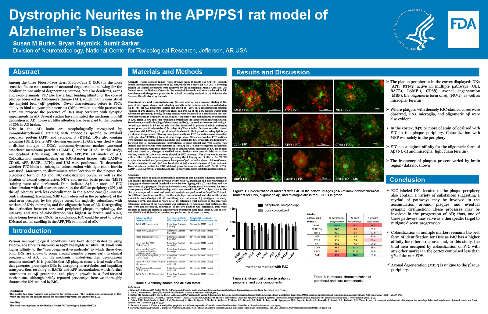
Among the three Fluoro-Jade dyes, Fluoro-Jade C (FJC) is the most sensitive fluorescent marker of neuronal degeneration, allowing for the localization not only of degenerating neurons, but also dendrites, axons and terminals. FJC also has a high binding affinity for the core of plaques observed in Alzheimer’s disease (AD), which mainly consists of the amyloid beta (A?) peptide. Never reported before is FJC’s ability to bind dystrophic neurites (DNs; swollen neuritic processes). Here, we propose the presence of DNs may correlate with synaptic impairments in AD. Several studies have indicated the mechanism of A? deposition in AD; however, little attention has been paid to the location of DNs in AD brains.
DNs in the AD brain are morphologically recognized by immunohistochemical staining with antibodies specific to amyloid precursor protein (APP) and reticulon 3 (RTN3). DNs also contain ubiquitin (UBI), ?-Site APP cleaving enzyme-1 (BACE1; enriched within a distinct subtype of DNs), endosome/lysosome marker lysosomal associated membrane protein 1 (LAMP-1), and/or CD68. In this study, we localized DNs using FJC in the APP/PS1 rat model of AD. Colocalization immunolabeling on FJC stained tissues with LAMP-1, CD-68, APP, BACE1, RTN3, and UBI were performed. To determine whether FJC binds to microglia, colocalization with light chain ferritin was used. Moreover, to discriminate if FJC primarily binds to the oligomeric form of A?, FJC colocalization with NU-1 and myelin basic protein (MBP) was also performed. Data indicate 85% or more of FJC colocalization with all markers occurs in the diffuse periphery of the A? plaques, with less colocalization in the plaque core (i.e.-intense central staining). However, excluding MBP (only observed in the periphery), of the total area occupied by the plaque cores, the majority colocalized with markers of DNs, microglia, and the oligomeric form of A?. Disregarding the distinction between core and peripheral plaque morphology, the intensity and area of colocalization was highest in ferritin and NU-1, while being lowest in CD68. In conclusion, FJC could be used to detect dystrophic neurites and axonal swelling in the APP/PS1 rat model of AD.