Impact Story: Improved Assessment of Cardiotoxic Risk in Drug Candidates: The Comprehensive in vitro Proarrhythmia Assay
A Spike in Drug-Induced Arrhythmias at the End of the Twentieth Century
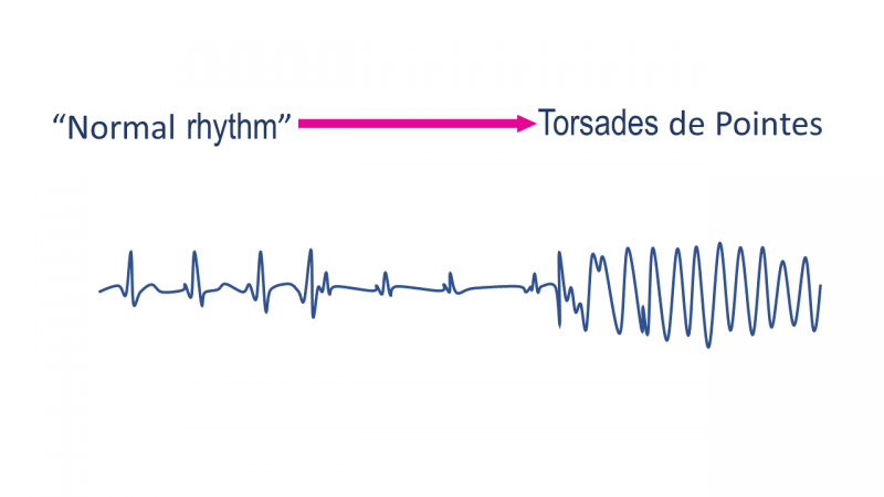
It has been nearly a century since doctors first reported that heart patients treated with quinidine, a derivative of quinine, could experience fainting spells, convulsions, and even fatal heart attacks. These patients showed “rhythms indicating intoxication of the heart muscle"1 and produced a distinctive electrocardiogram pattern that the French cardiologist Francois Dessertenne would later, in the 1960s, call torsades de pointes (TdP), meaning “twisting of the points.” TdP is now known to often lead to fatal heart arrhythmia.
Beginning in the late 1980s, cases of TdP in patients undergoing drug treatments increased markedly, leading to the withdrawal of several drugs from the U.S. and European markets. It became clear that the electrocardiograms of patients who experienced drug-associated TdP manifested a lengthened interval between the Q and T waves, compared to a typical heart beat; a longer QT interval was also observed in untreated individuals with a genetic predisposition to TdP called long QT syndrome. Surprisingly, the drugs linked to TdP were highly variable in their structures and their indications.
The Regulatory Response
In 2005, working with European and Japanese regulatory agencies through the International Committee for Harmonisation (ICH, now known as the International Council for Harmonisation), FDA participated in developing two key international regulatory guidances that outlined steps drug developers could take to determine if a new drug were associated with the risk of TdP. One guidance, focused on the preclinical setting, recommended testing the inhibitory effect of new drugs on the hERG protein, a voltage-gated potassium channel in cardiomyocytes that controls electrical activity of the heart. Mutations that affect the hERG protein have been associated with the development of TdP. The other guidance recommended that drug developers conduct randomized clinical trials in which the effects of the proposed new drug on the QT interval are measured.
After publication of the two guidances, no additional drugs with unanticipated TdP risk entered the market. Regulators, industry, and members of the academic community realized, however, that the strategies proposed by the guidance documents may have negatively impacted drug development, recognizing that although the preclinical and clinical tests outlined in the guidances could detect the potential to cause TdP, many of the estimated hundreds of drugs that interact with the hERG protein and prolong the QT interval do not cause torsades. Thus, decisions by developers to stop developing drugs with a hERG or QT signal could prevent promising drugs from reaching patients. For this reason, CDER investigators engaged in a sustained collaboration called CiPA (Comprehensive In Vitro Proarrythmia Assay), involving industry, the academic community, FDA, and regulatory agencies in Europe, Canada, and Japan, seeking to develop better ways to determine the potential of drugs to cause TdP.
Insights into the Mechanism of QT Prolongation and Torsades
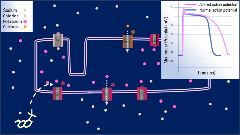
The heart’s function relies on the coordinated action of millions of rod-shaped muscle cells called cardiomyocytes that contract lengthwise with every heartbeat. Myocytes in each region of the heart must exert force at precisely the right moment if the heart is to pump blood efficiently. The exquisite timing of this contraction is controlled by the action potential, which is a periodic change in the cell’s electric potential over the course of a fraction of a second. The action potential is produced by the opening and closing of ion channel proteins that permit the movement of sodium, potassium, chloride, and calcium ions across the cell membrane. It is the changing intracellular concentrations of calcium ions that controls cardiomyocyte contraction.
The opening of the hERG channel to permit potassium ions to flow out of the cell plays a key role in repolarization, that is, bringing the membrane potential back to its resting state before the next round of depolarization. By blocking the hERG channel, many drugs can delay repolarization, and with this delay the cardiomyocyte is prone to activate and contract prematurely. Premature signal in a particular region of the heart can trigger irregular beats, including TdP.
The key insight as to how to develop a better biomarker for drug-induced TdP came from studies showing that many drugs simultaneously interact with other voltage-gated channels in addition to hERG and that these interactions may mitigate the risk of TdP due to hERG inhibition. For example, in an FDA-led investigation of 30 clinical drugs, it was observed that several low-risk drugs showed inhibition of calcium or sodium channels in addition to substantial hERG inhibition.2 These and other studies raised the possibility that one could develop biomarkers based on a more complete understanding of drug effects on voltage-gated channels that would be more specific than either QT prolongation in clinical studies or hERG inhibition in the laboratory.
Drug-induced torsades de pointes. The heart’s function depends on the coordinated contractions of individual muscle cells called cardiomyocytes. The timing of these contractions across the heart is controlled by a periodic electrical change in each cardiomyocyte (the action potential). The action potential is due to the controlled opening and closing of a variety of channels that are selective for different ions and allow or prevent their passage across the cell’s membrane. A variety of drugs, with very diverse structures, can interact with one or more of these channels and thereby alter the action potential to cause premature cardiomyocyte contraction and torsades de pointes, a potentially fatal form of arrythmia. The aim of the CiPA initiative is to accurately predict which drugs may cause torsades based on an understanding of their interactions with the various ion channels in the cardiomyocyte.
CiPA and Recent Advances in Determining Risk of Drug-Induced TdP
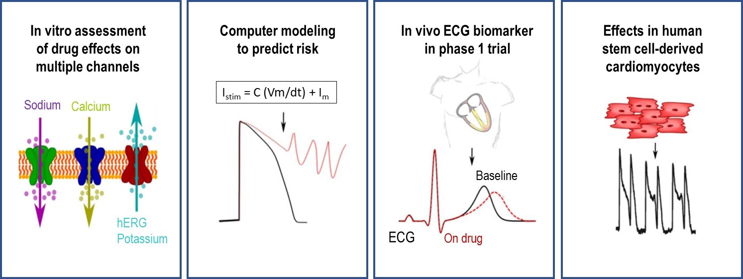
The CiPA (Comprehensive in vitro Proarrhythmia Assay) collaboration brings together FDA, its foreign counterparts (e.g., in Europe and Japan), non-profits, academia, and drug developers to better leverage indicators of the risk that a drug will cause TdP. CDER researchers and their CiPA partners have recently made important strides in each of the four components of the initiative:
- Assessment of drug effects on different ion channels. CDER investigators have identified essential criteria for methods and protocols with which developers could separately measure the interaction of a new drug molecule with different classes of cardiac ion channel using special cell lines developed in the laboratory.
- Computer modeling to predict risk based on drug effects on ion channels. Cellular assay data (component 1) on drug interactions with individual channels will be fed into a mathematical model of cardiomyocyte electrical signaling. Starting with a widely used model of the myocyte’s electrical activity, CiPA investigators have developed a sophisticated in silico approach to understand how voltage-gated channels and drugs that interact with them control the action potential.3 The in silico work has successfully conformed to a stringent and pre-specified validation strategy.
- Stem cell-derived Cardiomyocytes. Cardiomyocytes derived from pluripotent human stem cells are also proposed under the CiPA paradigm to confirm model-based predictions of the effects of new drugs and check for unanticipated effects. A recent multisite study by CDER investigators and their collaborators examined various responses of stem-cell-derived cardiomyocytes upon challenge by drugs with different TdP risks and alternative experimental approaches.4 The consistency of the results, obtained across multiple study sites and different experimental approaches, suggested that assays using these cells could be a very useful component of cardiac risk assessment in a sponsor’s drug development program.5
- Clinical testing. Clinical studies in the course of phase 1 trials would be used to check for any drug effects that were not predicted by preclinical testing. Recent work led by CDER investigators demonstrates that a new electrocardiogram measurement, the J-Tpeak, can be used to confirm the “balanced channel blocking” that has been associated with reduced TdP risk.6
The Implications of CiPA for Drug Development
Through its research to advance the four components of CiPA, CDER seeks to provide drug developers with the tools needed to better identify the risk of torsades posed by new drug compounds and to avoid the premature termination of promising candidates. In addition to being more specific for detecting cardiotoxic drugs, the emerging CiPA paradigm has the advantage of flexibility in that the ultimate amount of testing required is driven by the emerging data. As we look forward to fully implementing the paradigm, particular attention will be applied to developing more user-friendly experimental protocols to generate input data for the validated in silico model (components 1 and 2 above), developing standardized protocols for measuring the response of stem-cell derived myocytes to drug challenge (component 3), and further validating new electrocardiogram-based markers (component 4).
Despite the challenges, research advances like the ones outlined above have supported the formation of an international working group under the ICH7 to revise guidelines on how novel approaches can aid in determining the proarrhythmic risk of a drug and inform clinical development.8 The CiPA paradigm, promoting risk assessments that encompass multiple ion channels, is already being implemented for certain medications whose effects make it difficult to measure the QT interval.
The four components of the CiPA paradigm are individual assessment of drug effects on critical voltage-gated channels in vitro, computational modeling of the combined effects of drug-channel interactions on the cardiomyocyte action potential, and checking for unanticipated effects of drug-channel interactions using ECG-based biomarkers and stem cell-derived cardiomyocytes.
What are some of the potential benefits of the CiPA initiative for drug development? In developing better ways to predict the risk that a new drug molecule will cause dangerous arrythmias, CiPA has the potential to deter the inappropriate discontinuation of promising drugs and to provide more informative labels for new and currently marketed drugs.
[1] Levy Rl, Clinical studies of quinidin: The clinical toxicology of quinidin. JAMA.1922;79 (14):1108–1113.
[2] Crumb Jr, William J., et al. "An evaluation of 30 clinical drugs against the comprehensive in vitro proarrhythmia assay (CiPA) proposed ion channel panel." Journal of pharmacological and toxicological methods 81 (2016): 251-262.
[3] CDER investigators have elaborated the model of the cardiomyocyte to capture mechanistic details (e.g., the binding and unbinding of the drug to the hERG potassium channel over time). This model had increased power to discriminate drugs in different risk categories (Li and Strauss et al., Assessment of an In Silico Mechanistic Model for Proarrhythmia Risk Prediction under the CiPA Initiative. Clinical Pharmacology & Therapeutics. |https://doi.org/10.1002/cpt.1184).
[4] The two approaches used were microelectrode array or voltage sensing optical techniques.
[5] Blinova, Ksenia, et al. "International multisite study of human-induced pluripotent stem cell-derived cardiomyocytes for drug proarrhythmic potential assessment." Cell reports 24.13 (2018): 3582-3,
[6] Vicente, Jose, et al. "Assessment of Multi‐Ion Channel Block in a Phase I Randomized Study Design: Results of the CiPA Phase I ECG Biomarker Validation Study." Clinical Pharmacology & Therapeutics 105.4 (2019): 943-953.
[7] The International Council for Harmonisation (ICH), brings together representatives of the pharmaceutical industry and international regulatory authorities including the FDA to develop harmonised guidelines for global pharmaceutical development as well as their regulation.
[8] Commenting on the charge of the working group, the ICH website states “The objective of the first stage of the proposed harmonisation work is to provide clarity on how to standardise assays such as multi-ion channel assays, in silico models, in vitro human primary and induced pluripotent cardiomyocyte assays and in vivo evaluation, and apply these learnings to guide predictions and subsequent clinical assessment. These efforts will provide a customisable non-clinical strategy that is more informative for clinical development.”


